Free Online Assessment
 WHATSAPP
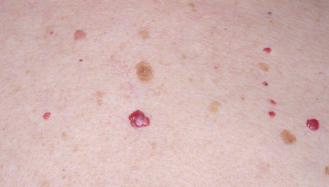
Cherry angiomas often appear suddenly on the skin, causing concern for many people. These small, bright red spots are usually harmless but can sometimes signal underlying health issues. Understanding why they develop and when they warrant medical attention can help you manage these common skin growths effectively and with peace of mind.
- Dr Sharon Crichlow
- Reading Time: 10 Mins
Cherry angiomas often appear suddenly on the skin, causing concern for many people. These small, bright red spots are usually harmless but can sometimes signal underlying health issues. Understanding why they develop and when they warrant medical attention can help you manage these common skin growths effectively and with peace of mind.
Key Takeaways
- Cherry angiomas are benign red skin spots caused by clustered blood vessels, commonly appearing after age 30 and increasing with age.
- While usually harmless, sudden eruption of multiple angiomas could signal underlying health conditions requiring medical evaluation.
- Professional removal methods like laser therapy or cryotherapy are safe and effective if angiomas cause cosmetic concerns or discomfort.
Table of Contents
What Are Cherry Angiomas?
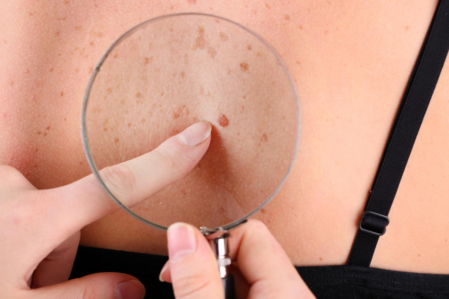
Cherry angiomas are small, round skin growths that appear as bright red dots on the skin. They’re made up of clusters of dilated blood vessels, giving them their distinctive red appearance.
These benign growths typically range from 1-5mm in diameter and can be flat or slightly raised. Most people develop them after age 30, with prevalence increasing significantly with age. Research shows approximately 75% of adults over 75 years have at least a few cherry angiomas.
The technical name for these growths is Campbell de Morgan spots, though they’re also sometimes called senile angiomas or ruby spots. Unlike some skin conditions, cherry angiomas don’t typically cause pain, itching, or discomfort.
Cherry angiomas most commonly appear on the trunk, especially the chest and back, but can develop anywhere on the body. They’re permanent once they appear, though their size and number may increase over time.
| Characteristic | Description |
|---|---|
| Size | 1–5mm in diameter |
| Colour | Bright red to purple |
| Texture | Smooth, dome-shaped |
| Feel | Painless unless irritated |
| Common Locations | Torso, arms, legs |
Why Cherry Angiomas Appear Suddenly
The exact cause of cherry angiomas remains incompletely understood, but several factors have been linked to their sudden appearance:
Genetic Factors
Research has identified specific genetic mutations associated with cherry angiomas. Studies published in dermatology journals have found somatic missense mutations in genes called GNAQ and GNA11 (particularly Q209H) in many cherry angiomas.
These genetic findings suggest that cherry angiomas represent clonal vascular proliferation rather than simple degenerative changes, explaining why they can sometimes appear suddenly in clusters.
Age-Related Changes
Age remains the strongest predictor for developing cherry angiomas. The incidence increases dramatically after age 30, suggesting that cellular and vascular changes that occur with ageing play a significant role in their formation.
A cross-sectional analysis conducted by Borghi et al. at the University of Ferrara examined 1,302 dermatology patients and confirmed advanced age as a significant risk factor for eruptive cherry angiomas (defined as having 30 or more lesions).
Medical Conditions
Several medical conditions have been associated with sudden eruptions of cherry angiomas:
- Metabolic disorders: A study found that patients with Type 2 diabetes had significantly more cherry angiomas than non-diabetic controls, suggesting that metabolic stress may trigger their formation.
- Immunosuppression: The Borghi study identified chronic iatrogenic immunosuppression as the strongest factor associated with eruptive cherry angiomas, even stronger than age. They hypothesised that an imbalance in skin immune competence might predispose to angioma formation.
- Malignancies: Multiple studies have found associations between cherry angiomas and various malignancies, especially skin tumours.
- Lymphoproliferative disorders: A case reported in JAMA Dermatology described a 25-year-old male who experienced the sudden appearance of 23 cherry angiomas over just 5 days, which turned out to be a presenting sign of multicentric Castleman disease.
Chemical and Environmental Factors
Environmental exposures have been documented as triggers for cherry angiomas:
- Chemical exposure: Associations have been observed between cherry angiomas and exposure to certain chemicals, including bromides, sulfur mustard, and butoxyethanol.
- Medications: Nazer’s study found that tamsulosin use was significantly associated with increased cherry angioma risk, while clopidogrel use appeared protective. They speculated this might relate to the vascular effects of these medications.
Hormonal Changes
Hormonal fluctuations, particularly during pregnancy, have been linked to the sudden appearance of cherry angiomas. Some research suggests that elevated oestrogen levels may stimulate angiogenesis (blood vessel formation), contributing to their development.
Warning Signs to See a Doctor
While most cherry angiomas are harmless, certain situations warrant medical evaluation.
The rapid development of numerous cherry angiomas (sometimes called “eruptive cherry angiomas”) can be concerning. Studies suggest this might indicate underlying conditions like lymphoproliferative disorders or systemic disease.
If you notice a sudden explosion of multiple cherry angiomas over days or weeks, consult a dermatologist for proper evaluation.
Moreover, any significant changes to existing cherry angiomas should prompt medical attention.
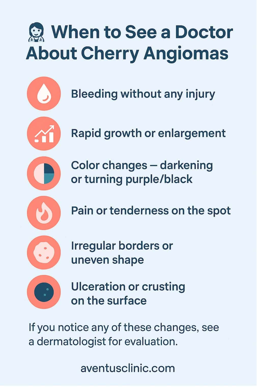
These changes could potentially indicate a different type of skin lesion that requires proper diagnosis.
Treatment and Removal Options
Most cherry angiomas don’t require treatment unless they cause physical discomfort or cosmetic concerns. Several effective removal methods exist:
Professional Removal Methods
Electrocautery: Uses heat to burn and destroy the angioma. This is quick and effective for smaller angiomas.
- Cryotherapy: Freezes the angioma with liquid nitrogen, causing the tissue to die and fall off.
- Laser therapy: Particularly effective for cherry angiomas, lasers target the blood vessels without damaging surrounding skin.
- Shave excision: For larger angiomas, this method involves shaving the growth off at skin level.
| Removal Method | Best For | Recovery Time | Potential Side Effects |
|---|---|---|---|
| Electrocautery | Small, individual angiomas | 1–2 weeks | Slight scarring, temporary discolouration |
| Cryotherapy | Surface angiomas | 1–3 weeks | Temporary blister, possible hypopigmentation |
| Laser Therapy | Multiple angiomas, sensitive areas | 1–2 weeks | Mild redness, rarely scarring |
| Shave Excision | Larger angiomas | 2–3 weeks | Potential scarring, infection risk |
Professional treatment ensures proper technique and reduces risks of scarring or infection. Our clinic specialises in the removal of various skin lesions, including cherry angiomas.
Home Care Approaches
While professional removal is recommended, some people try home approaches to reduce the appearance of cherry angiomas:
- Topical retinoids: May help reduce the appearance over time, but won’t eliminate them completely.
- Apple cider vinegar: Sometimes applied to dry out small angiomas, though medical evidence for effectiveness is lacking.
- Tea tree oil: Has anti-inflammatory properties but minimal evidence for cherry angioma treatment.
Important: Never attempt to cut, pick, or remove angiomas at home as this can lead to infection, scarring, and excessive bleeding.
Monitoring and Self-Care
If you have cherry angiomas, regular self-examination helps track any changes.

Cherry angiomas rarely cause complications, but keeping them clean and protected from injury helps prevent potential issues like infection or excessive bleeding.
Conclusion
Cherry angiomas are generally harmless, but understanding when they might signal something more serious is important for your health. If you’re concerned about sudden or unusual cherry angiomas, professional evaluation can provide peace of mind and treatment options if desired. For personalised advice about cherry angiomas or other skin concerns, complete our free online assessment to speak with our skin specialists.
FAQs
Can cherry angiomas appear suddenly without warning?
Yes, cherry angiomas can appear suddenly, sometimes in clusters. This may be related to age, hormonal changes, genetics, or rarely, underlying medical conditions.
Are sudden multiple cherry angiomas a sign of a serious health problem?
While most cases are benign, the sudden appearance of numerous cherry angiomas should be evaluated by a doctor, especially if accompanied by other symptoms like weight loss, fatigue, or enlarged lymph nodes.
Can hormones or medications trigger sudden cherry angiomas?
Yes, hormonal changes (especially during pregnancy) and certain medications like tamsulosin have been associated with increased cherry angioma formation.
Do cherry angiomas ever bleed or cause pain?
Cherry angiomas don’t typically cause pain but can bleed profusely if injured due to their high concentration of blood vessels. Normal cherry angiomas shouldn’t bleed spontaneously.
How can I safely remove cherry angiomas if they are cosmetically concerning?
Professional removal through laser therapy, electrocautery, or cryotherapy is safest and most effective. Home removal attempts can lead to infection, scarring, and excessive bleeding.