

Red moles are typically harmless cherry angiomas, but could be melanoma. Cherry angiomas are bright red, round, and stable. Melanoma shows irregular borders, colour variations, and rapid growth. In rare cases, a rapidly growing red or purple nodule may represent Merkel cell carcinoma, an uncommon but aggressive skin cancer. Consult a doctor for concerning changes.
- Dr Sharon Crichlow
- Reading Time: 10 Mins
Spotting a bright red mole on your skin can be worrying. While most red moles are harmless cherry angiomas, some could be signs of skin cancer. In uncommon cases, red or pink lesions may represent amelanotic melanoma or, rarely, Merkel cell carcinoma. Knowing the difference between these common skin growths and potentially dangerous melanoma could save your life. Let’s explore how to identify these skin lesions and when you should seek medical advice.
Key Takeaways
- Cherry angiomas are benign red moles that remain stable, while melanomas show irregular growth and changes.
- Use the ABCDE rule (Asymmetry, Border, Colour, Diameter, Evolution) to identify suspicious red skin lesions.
- Rapidly enlarging, firm red nodules in older adults should also be assessed to exclude rare conditions such as Merkel cell carcinoma.
- See a dermatologist for any red moles that bleed unexpectedly, change appearance, or grow rapidly.
Table of Contents
What causes a red mole on the skin?
It is common to notice a small red mole on the skin and feel concerned. Any new or changing lesion can raise questions about skin cancer. In most cases, however, a bright red mole is a cherry angioma, a benign growth of small blood vessels.
Melanoma, a type of skin cancer arising from pigment-producing cells, can sometimes appear red or pink rather than dark brown or black. In addition, Merkel cell carcinoma, a rare neuroendocrine skin cancer, may present as a red or violaceous lump that grows quickly. Because of this, distinguishing between harmless vascular growths and potentially serious lesions is important.
Understanding how cherry angiomas form, how melanoma behaves, and what warning signs to look for can help you decide when reassurance is appropriate and when medical review is necessary.
Medical Disclaimer: This content is for general informational purposes only and does not constitute medical advice. It should not replace professional medical consultation, diagnosis or treatment. If you are concerned about a red mole or notice any changes in your skin, seek assessment from a qualified healthcare professional.
What is a cherry angioma?
A cherry angioma is a benign vascular skin growth made up of clusters of dilated capillaries. These small blood vessels enlarge and group together near the surface of the skin, creating a visible red or purple spot.
Cherry angiomas are:
- Non-cancerous
- Not contagious
- Not caused by infection
- Not related to melanoma
They are one of the most common benign skin growths in adults. Their exact cause is not fully understood, but they are strongly associated with ageing. Most people begin developing them after the age of 30, and the number often increases over time.
Hormonal influences, genetic predisposition and certain environmental exposures have been suggested as contributing factors, but no single definitive cause has been established.
Importantly, cherry angiomas do not turn into melanoma.
What causes cherry angiomas to form?
Cherry angiomas develop when tiny blood vessels in the skin dilate and proliferate. This creates a small cluster of capillaries that becomes visible through the surface of the skin.
They are more common:
- In adults over 30
- During pregnancy
- In individuals with a family history of similar lesions
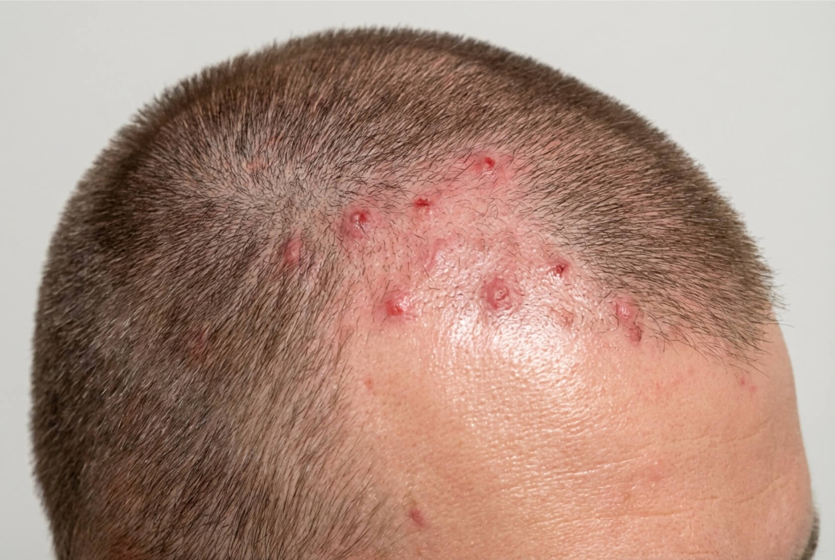
In rare cases, a sudden eruption of multiple cherry angiomas has been associated with underlying medical conditions or exposure to certain chemicals, but this is uncommon.
Most cherry angiomas develop gradually and remain stable.
What does a cherry angioma look like?
Cherry angiomas have distinct clinical features:
| Feature | Description |
|---|---|
| Colour | Bright red, ruby-red, or purple-red |
| Shape | Round or oval, dome-shaped |
| Size | Usually small (1–5mm in diameter) |
| Texture | Smooth, often slightly raised |
| Border | Well-defined, clear edges |
| Surface | May appear as a collection of tiny blood vessels |
They are typically painless. Because they are made up of blood vessels, they may bleed if scratched or traumatised.
A study of dermoscopic features of cherry angiomas showing that a large proportion of lesions demonstrate typical lacunae, with red to red-blue shades, on dermoscopic examination. Under dermoscopy, cherry angiomas often display characteristic red or purple “lacunar” patterns, which help dermatologists differentiate them from malignant lesions.
They most commonly appear on the trunk, particularly the chest and back, but can develop anywhere on the body.
What does melanoma look like on the skin?
Melanoma typically appears as an irregular, dark-coloured mole. However, amelanotic melanoma, a less common variant, can appear reddish, pink, or flesh-coloured, making it easily confused with cherry angiomas.
Warning signs of melanoma follow the ABCDE rule:
- Asymmetry: Uneven shape
- Border: Irregular, ragged, or blurred edges
- Colour: Varied shades within the same lesion
- Diameter: Larger than 6mm (though can be smaller)
- Evolving: Changes in size, shape, or colour
If you’re concerned about any moles on your skin, our comprehensive mole checking service provides thorough evaluations with same-day results in most cases.
Could a red mole be merkel cell carcinoma?
Merkel cell carcinoma (MCC) is a rare but aggressive type of skin cancer. It arises from neuroendocrine cells in the skin and most commonly affects older adults.
Unlike melanoma, MCC does not follow the ABCDE rule. Instead, clinicians often refer to the AEIOU features:
- Asymptomatic (usually painless)
- Expanding rapidly
- Immune suppression
- Older than 50
- UV-exposed skin
Merkel cell carcinoma typically appears as:
- A firm, dome-shaped nodule
- Red, pink, purple, or skin-coloured
- Rapidly enlarging over weeks to months
- Smooth and shiny in appearance
- It most often develops on sun-exposed areas such as the head, neck, and upper limbs.
Although rare, any rapidly growing, firm red or violaceous nodule should be assessed promptly.
If you’re concerned about any moles on your skin, our comprehensive mole checking service provides thorough evaluations with same-day results in most cases.
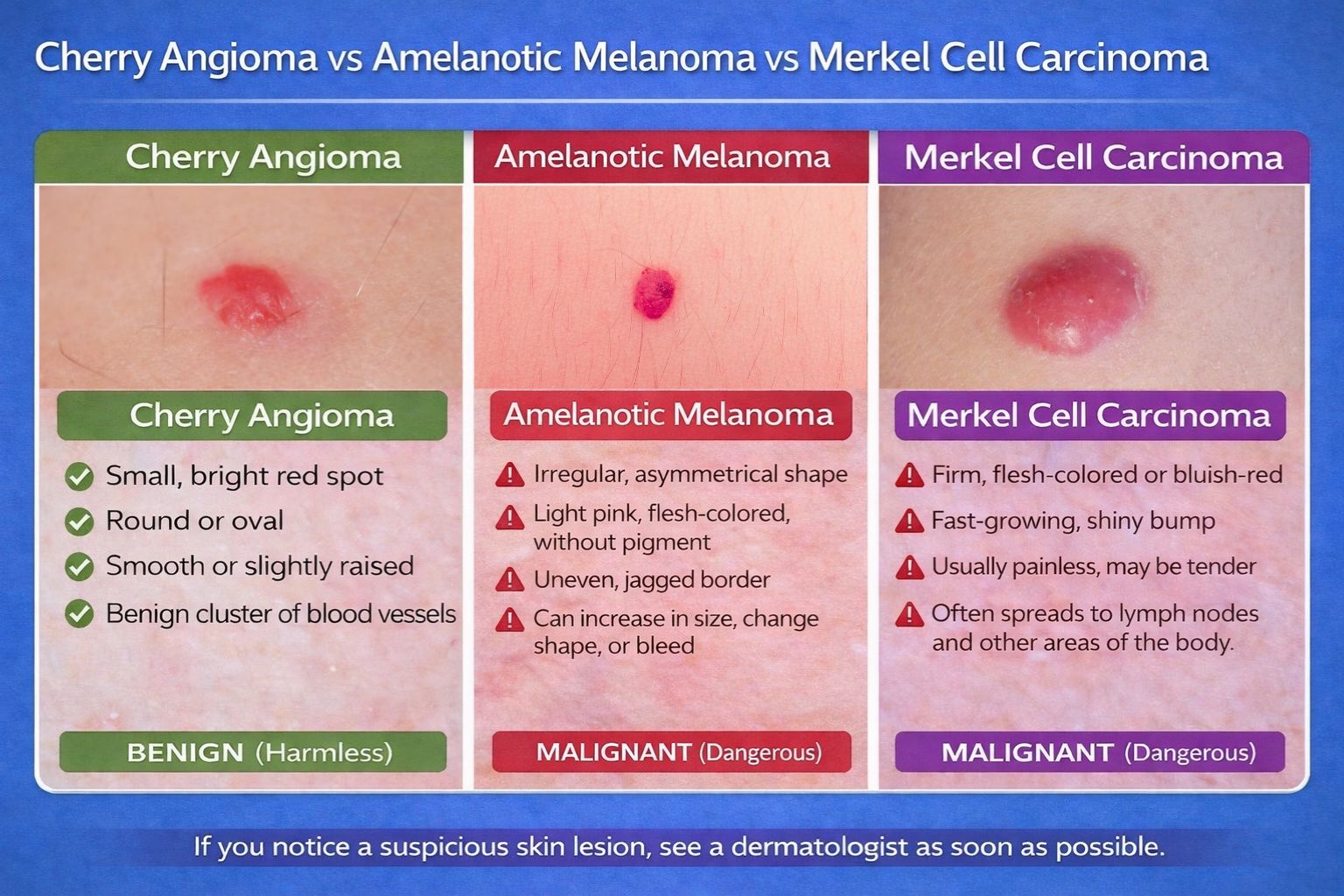
Cherry angioma vs. melanoma vs. merkel cell carcinoma: How to spot the difference
Distinguishing between these two conditions is crucial:
| Feature | Cherry Angioma | Melanoma | Merkel Cell Carcinoma |
|---|---|---|---|
| Colour Pattern | Uniform red/purple | Often varied | Red, pink, purple or skin-coloured |
| Border | Well-defined, smooth | Irregular or blurred | Usually smooth but rapidly enlarging |
| Surface | Smooth, dome-shaped | May be uneven or ulcerated | Smooth, firm nodule |
| Growth | Slow, minimal change | May grow or change | Rapid enlargement |
| Vascular Pattern | Red-purple lacunae | Polymorphous/irregular vessels | No lacunar pattern |
| Feel | Soft | Variable | Firm |
| Age of Onset | More common after 30 | Can appear at any age | Usually over 50 |
If you’re uncertain about a red lesion, it’s always best to consult a specialist dermatologist for professional evaluation and peace of mind.
Common areas where red moles appear
Cherry angiomas can develop anywhere on the body, but certain locations are more common. A population imaging study published in Dermatology (2022) mapped their distribution, showing a clear preference for the trunk.
The chest and back are prime locations, followed by the arms and shoulders. They’re less common on the face, hands, and feet. This distribution pattern can help distinguish them from other skin conditions.
Location alone doesn’t determine whether a growth is concerning, but it provides helpful context. Cherry angiomas on the face or neck may cause more cosmetic concerns, while those under clothing might experience irritation.
Any new or changing growth deserves attention, regardless of location. The most important factor is how the growth behaves over time.
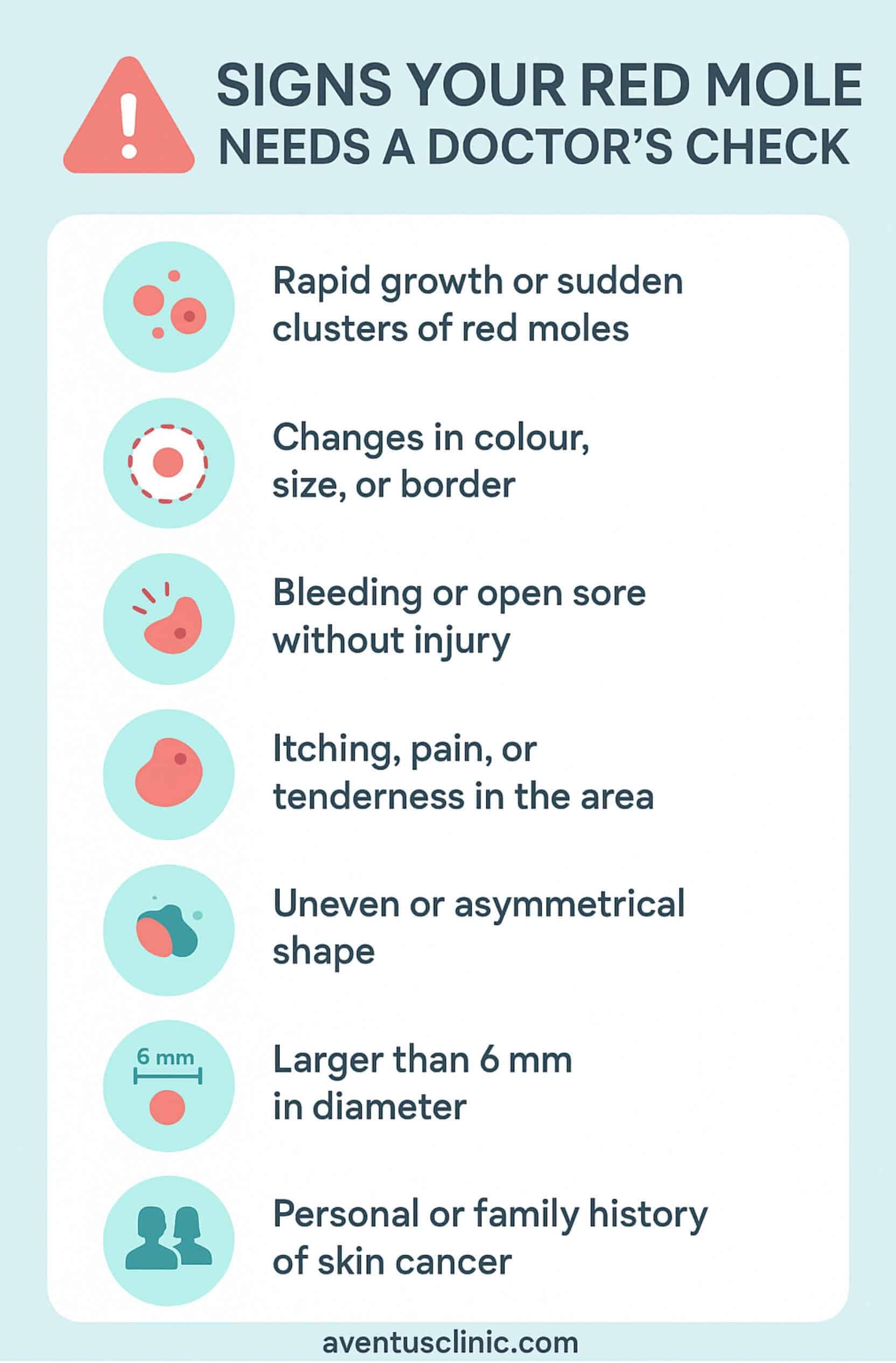
When should you see a dermatologist for a red mole?
While most red moles are harmless, certain warning signs should prompt medical evaluation.
A study noted that eruptive cherry angiomas (sudden appearance of multiple lesions) showed an association with malignant melanoma, suggesting that patients with this presentation merit careful skin examination.
Trust your instincts. If something doesn’t seem right about a skin lesion, seeking professional evaluation is always the safer choice.
How are red moles diagnosed and treated?
Proper diagnosis of red skin lesions relies on several advanced techniques.
The diagnostic process for red moles typically begins with a thorough visual examination. Your dermatologist will first assess the growth with the naked eye, then use a dermatoscope for more detailed viewing.
If there’s any uncertainty, a biopsy may be recommended. This involves removing all or part of the growth and examining it under a microscope.
For confirmed cherry angiomas, several treatment options exist:
| Treatment | How It Works | Recovery Time |
|---|---|---|
| Laser Removal | Uses targeted light energy to collapse blood vessels | Minimal downtime, slight redness for 1–2 days |
| Cryotherapy | Freezes the growth with liquid nitrogen | Blister forms and heals within 1–2 weeks |
| Electrocautery | Uses electrical current to heat and remove tissue | Small scab forms and falls off within 1–2 weeks |
Cherry angioma removal is typically cosmetic. Melanoma and Merkel cell carcinoma require urgent specialist treatment to prevent spread.
Our clinic provides safe and effective mole removal in Hertfordshire for both cosmetic and medical concerns, with histological analysis of all removed tissue.
Conclusion
While most red moles are harmless cherry angiomas, a small proportion may represent amelanotic melanoma or, rarely, Merkel cell carcinoma. Regular skin checks and prompt medical attention for suspicious or rapidly changing lesions are essential steps in skin cancer prevention. If you’re concerned about a red mole, start with our free online skin assessment to get expert guidance on the next steps for your skin health.
FAQs
Can a red mole appear suddenly and still be harmless?
Yes. Cherry angiomas can develop quite suddenly and still be completely benign. However, the rapid appearance of multiple red moles or dramatic changes in existing ones should be evaluated by a dermatologist.
Are cherry angiomas linked to stress or hormones?
Hormonal changes, particularly during pregnancy, can trigger the development of cherry angiomas. While stress isn’t directly linked, some patients report new angiomas during high-stress periods, though scientific evidence for this connection is limited.
Can melanoma start as a red mole?
Yes. Amelanotic melanoma often appears as a red, pink, or skin-coloured lesion rather than the typical dark mole. Any red lesion that changes, grows, bleeds, or has irregular borders should be examined promptly.
Do cherry angiomas go away on their own?
Cherry angiomas don’t typically disappear once they’ve formed. They may grow slightly larger with time, but generally remain stable throughout life unless treated or removed.
Should I remove a red mole for cosmetic reasons?
Removing cherry angiomas for cosmetic reasons is safe and common. Professional removal offers better cosmetic results and allows for histological examination if there’s any uncertainty about the diagnosis.

Concerned about a red mole on your skin? Get expert advice from our dermatology specialists with a free online assessment.

 CALL US
 WHATSAPP