Overview
What are epidermoid cysts?
Pilar cysts are non-cancerous and usually slow growing. They account for a significant proportion of cysts found on the scalp and are most often seen in middle-aged adults. Women are affected slightly more often than men. A family history is common, and in some individuals pilar cysts follow an autosomal dominant inheritance pattern, meaning they can run in families.
Although most are harmless, any new or changing lump should be assessed to confirm the diagnosis.
Size and appearance
Pilar cysts typically measure between 5 millimetres (0.5 cm) and 5 centimetres in diameter. Many are around 1 to 3 centimetres in size. They often begin small, approximately 5 to 10 millimetres, and enlarge gradually over months or years. Larger cysts exceeding 5 centimetres are less common but can occur.
They are usually:
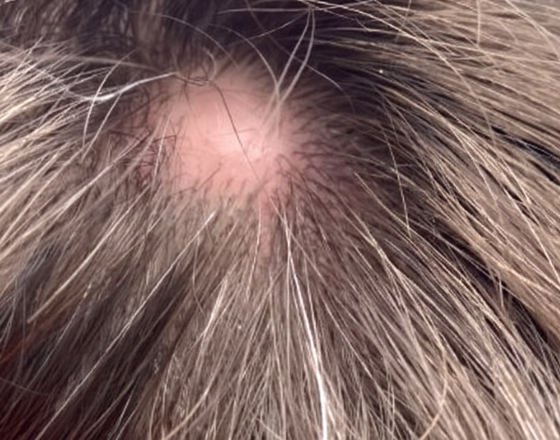
- Round or dome-shaped
- Firm and smooth
- Freely movable under the skin
- Covered by normal-appearing skin
Unlike epidermoid cysts, pilar cysts do not usually have a visible central punctum. Although most develop on the scalp, they can occasionally appear on the face, neck or trunk.
Symptoms and Causes
What causes a pilar cyst?
Pilar cysts form when cells from the outer root sheath of a hair follicle multiply and produce keratin, the structural protein found in hair and the outer layer of skin. Instead of shedding normally, keratin becomes trapped beneath the skin, forming a cyst wall and filling the cavity with compact keratin.
The exact trigger is not always known. Contributing factors may include:
- Genetic predisposition
- Familial inheritance
- Abnormal follicular keratinisation
Trauma is not considered a primary cause.
What symptoms do pilar cysts cause?
Most pilar cysts are painless and cause no symptoms other than a visible or palpable lump.
Symptoms may occur if the cyst becomes inflamed, ruptures or becomes infected. Signs of inflammation or infection can include redness, tenderness, swelling, warmth or discharge of thick keratinous material. Rapid enlargement, increasing pain or persistent inflammation should be medically assessed.
Diagnosis and Tests
How is a pilar cyst diagnosed?
Diagnosis is usually clinical. A GP or dermatologist can often identify a pilar cyst based on its typical features, particularly its location on the scalp, its firm and smooth structure, its mobility under the skin and the absence of a central punctum.
If there is diagnostic uncertainty, rapid change, atypical features or concern about malignancy, the cyst may be surgically removed and sent for histological examination.
Microscopic examination confirms the diagnosis by identifying:
- A cyst wall without a granular layer
- Dense, compact keratin within the cavity
Malignant transformation is rare but has been described in uncommon cases as proliferating trichilemmal tumours, which require specialist assessment.
Management and Treatment
Do pilar cysts need treatment?
Treatment is not always necessary. Many pilar cysts can be safely left alone if they are small, asymptomatic and not cosmetically concerning.
When is removal recommended?
Removal is not routinely required for pilar cysts. However, surgical excision may be advised if the cyst:
- Becomes painful, tender or persistently inflamed
- Repeatedly ruptures or leaks keratin material
- Develops signs of infection, such as redness, warmth or swelling
- Continues to enlarge over time
- Causes cosmetic concern or psychological distress
- Interferes with brushing, washing or routine hair care
Excision is usually performed under local anaesthetic and is most effective when the entire cyst wall is removed, which reduces the likelihood of recurrence.
How are pilar cysts removed?
The definitive treatment is complete surgical excision for pilar cyst, usually performed under local anaesthetic. Removing the entire cyst wall reduces the risk of recurrence.
If a cyst is inflamed or infected, management may include antibiotics where bacterial infection is present and delayed excision once inflammation has settled.
Incision and drainage alone does not remove the cyst wall and may lead to recurrence.
Outlook/Prognosis
The outlook for pilar cysts is excellent. They are benign, non-cancerous and slow growing.
After complete excision, recurrence is uncommon but can occur if part of the cyst lining remains. Individuals with a hereditary tendency may develop multiple cysts over time.
Having a pilar cyst does not increase the risk of skin cancer. However, any cyst that changes rapidly, ulcerates or behaves unusually should be reviewed by a clinician.
Prevention
There is no proven way to prevent pilar cysts, particularly when they are genetically determined. Because many cases have a hereditary basis, prevention is limited. Routine scalp hygiene does not prevent cyst formation. Early assessment of new or changing lumps allows appropriate diagnosis and reassurance.