

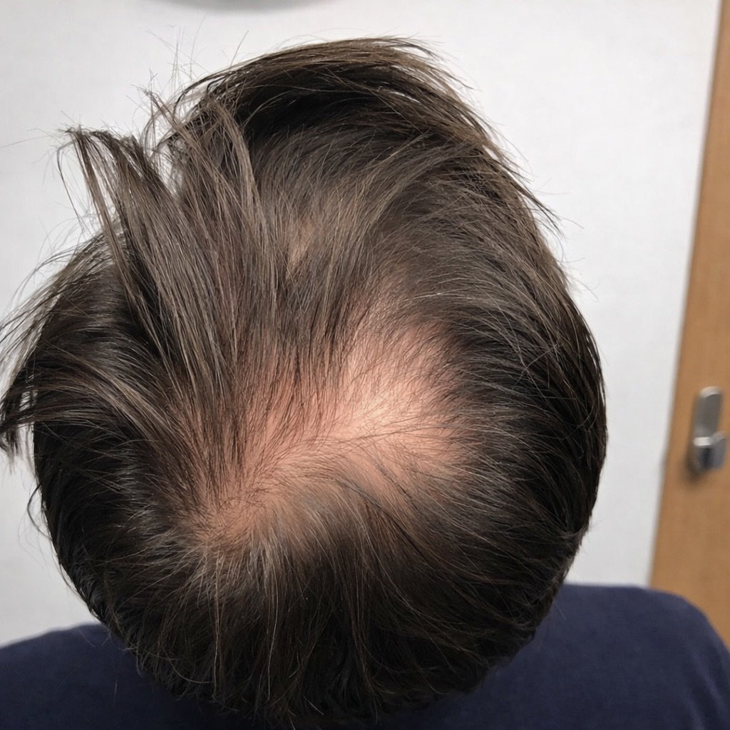
The crown often appears thinner under bright overhead lighting because light reflects off the scalp surface, and hair grows in a spiral pattern that naturally exposes more scalp area.
The crown can appear bald under bright overhead lighting or camera flash even without hair loss. Light reflects off scalp skin, the natural hair whorl creates spacing, and certain angles expose thinner coverage. Distinguishing optical illusion from follicle miniaturisation helps determine when assessment is needed.
Key Takeaways
- Crown hair grows in spiral whorls that naturally separate strands, making scalp more visible under direct overhead lighting.
- Follicle miniaturisation gradually reduces hair shaft diameter and density, increasing scalp show through beyond normal lighting effects.
- Progressive crown thinning with finer hair strands signals pattern hair loss requiring medical evaluation and possible treatment intervention.
Why the crown hair may look bald under bright lighting
The crown often appears thinner under direct or overhead light due to scalp reflection and natural hair growth patterns, rather than immediate hair loss affecting follicle health. Several visual and biological factors can make the crown appear sparse under bright overhead lighting.
Let’s explore the main factors and why they influence scalp visibility.
Light reflection increases scalp visibility
Bright overhead lighting reflects off the scalp surface and makes gaps between individual hairs easier to see compared to softer diffuse illumination.
Optical studies examining skin and hair under different illumination conditions show that angular lighting significantly affects scalp visibility because the scalp surface scatters light differently than hair shafts. It creates contrast that makes spacing more noticeable under direct sources positioned above the head.
Crown hair frows in a spiral pattern
Hair whorls at the crown create spiral growth patterns that radiate outward from a central point.
This arrangement separates hairs more than parallel growth elsewhere on the scalp, so gaps appear larger from above. The whorl centre often shows more scalp even when the density remains normal.
Perceived thinning versus real hair loss
Lighting and styling can create the illusion of thinning even when hair coverage remains normal. True hair loss occurs when follicles gradually produce finer, shorter hairs over time. Distinguishing optical effects from genuine miniaturisation requires consistent observation under normal lighting rather than relying on single photos.
Why hair density varies across the scalp
Hair density, shaft diameter, and follicular grouping vary naturally across different scalp regions and between individuals regardless of hair loss status.
Research measuring hair fibre characteristics across populations documents significant variation in diameter, pigmentation, and structural properties that affect coverage appearance. Crown areas typically show slightly lower density in many people compared to temporal or occipital regions.
Several biological characteristics determine how well hair covers the scalp, even when overall density appears similar. Some of those factors include:
- Follicles per square centimetre often range between about 100 and 200, depending on genetics and ethnicity
- Hairs per follicular unit typically average 1 to 4 strands emerging from each follicle opening
- Hair shaft thickness commonly varies between roughly 50 and 100 micrometres in diameter
- Hair pigmentation influences visibility because darker hair usually provides stronger visual scalp coverage
Together, these factors influence how much scalp becomes visible, particularly under bright overhead lighting.
Early crown thinning caused by follicle miniaturisation
Progressive follicle miniaturisation drives pattern hair loss by altering hair cycle dynamics so follicles produce thinner, shorter hairs with each successive growth phase. This mechanism increases scalp show through, especially under bright direct lighting that highlights gaps between thinner strands.
The following comparison shows how healthy follicles differ from miniaturised ones in crown thinning progression:
| Characteristic | Healthy Terminal Hair | Miniaturized Hair |
|---|---|---|
| Shaft diameter | 70 to 100 micrometres | 30 to 60 micrometres |
| Anagen phase duration | 2 to 7 years | 6 months to 2 years |
| Hair length potential | Long enough for coverage | Short, fails to cover scalp |
| Visual coverage | Dense scalp concealment | Increased light penetration |
| Dermoscopy appearance | Uniform thick shafts | Mixed thick and thin hairs |
Progressive follicle miniaturisation drives pattern hair loss by reducing hair shaft diameter and overall density with each growth cycle.
Dermoscopy and trichoscopy reveal hair diameter diversity where thick terminal hairs mix with progressively thinner vellus-like hairs, creating visible contrast that indicates ongoing miniaturisation requiring clinical assessment.
The role of DHT in crown hair loss
Dihydrotestosterone shortens the hair growth cycle and shrinks genetically susceptible follicles through androgen receptor binding that triggers miniaturisation cascades.
The crown and temples contain the highest concentration of androgen-sensitive follicles, which explains why vertex pattern hair loss commonly presents as isolated balding patches at the crown while occipital and lateral regions maintain normal density.
DHT gradually turns thick terminal hairs into fine vellus hairs over multiple growth cycles, reducing coverage and making the crown appear progressively balder under all lighting conditions rather than just harsh overhead sources.
Other factors that can make the crown look thinner
Several non-disease factors affect how crown coverage appears under different lighting and styling conditions beyond actual follicle miniaturisation. These often combine to create temporary appearance changes that improve with simple adjustments rather than requiring medical intervention.
Here is how common non-medical factors affect crown appearance and what corrective actions help improve visual coverage:
| Factor | Why It Affects Appearance | What Helps |
|---|---|---|
| Fine or light coloured hair | Reflects more light from scalp surface | Use darker products or temporary colour |
| Oil, sweat, or product buildup | Causes clumping that separates strands | Regular shampooing, avoid heavy oils |
| Styling that flattens crown | Removes natural volume helping coverage | Volumising products, blow-dry with round brush |
| Photographic angle and flash | Creates harsh overhead lighting | Use diffuse natural light, forward angles |
| UV photodamage to hair | Alters protein structure and light scattering | Protect hair with UV spray, wear hats |
Long term UV exposure chemically alters hair proteins, including keratin and melanin, changing surface properties and light scattering behaviour that affects how hair appears under illumination.
These environmental and styling factors distinguish temporary appearance issues from genuine follicle miniaturisation requiring medical treatment.
How doctors evaluate crown hair Tthinning
Clinical assessment combines patient history, visual examination, and specialised imaging to distinguish normal variation from progressive miniaturisation requiring treatment.
Clinical scalp examination and patient history
Doctors assess crown thinning through clinical scalp examination and patient history. They review when changes began, family history of pattern hair loss, medications, and recent stressors.
Physical examination evaluates hair density distribution, scalp inflammation or scaling, and gentle hair pull tests for excessive shedding.
Dermatoscopy and density assessment
Trichoscopy helps detect miniaturised hairs through magnified scalp examination, showing mixed hair diameters in affected areas.
Clinicians also assess density using phototrichograms or handheld dermoscopy devices, which magnify the scalp surface and allow doctors to evaluate follicle activity and hair growth patterns during consultation.
Treatment options for crown thinning
Medical, topical, device-based, and surgical interventions address crown thinning through different mechanisms depending on severity and patient preferences.
Topical minoxidil
Applied twice daily, minoxidil prolongs anagen phase and increases follicle size, slowing miniaturisation progression and maintaining existing density when started early before significant follicle atrophy occurs.
Oral finasteride or dutasteride
These medications block DHT conversion and reduce androgen effects on susceptible follicles. They suit only men due to pregnancy risks for women of childbearing age and require continuous use to maintain benefits.
Low-level laser and LED therapy
Device-based treatments deliver red light that stimulates cellular metabolism and may produce modest density improvements over six to twelve months of consistent regular use at home.
Hair transplant surgery
Surgical restoration relocates follicles from permanent zones to thinning crown areas. Crown restoration requires careful planning because the curved surface and natural whorl patterns demand artistic placement for natural-looking results.
Cosmetic and styling approaches
Fibre powders, scalp concealers, and thickening shampoos provide immediate visual improvement while medical treatments work gradually over months. These temporary solutions help boost confidence during treatment phases.
Early treatment preserves more follicles because miniaturisation becomes harder to reverse as follicles shrink toward complete atrophy.
For personalised treatment plans, we offer crown restoration treatments where we explain medication options, procedural interventions, and realistic outcome expectations based on your specific pattern and progression stage.
Conclusion
If your crown looks more visible under harsh lighting, it often reflects the lighting angle, natural hair whorl anatomy, and optical effects rather than immediate hair loss. Photograph your crown monthly under consistent conditions to track changes. If thinning progresses with finer strands, we can examine your crown and recommend appropriate treatment options.
Get a free quote and book a consultation today.
FAQs
Is it normal to see scalp at the crown?
Yes, because hair grows in whorls that naturally separate strands at the crown centre, making scalp more visible under bright overhead lighting even with normal density.
Why does my crown look bald in photos but not in mirror?
Camera flash creates harsh overhead lighting and angles that exaggerate natural spacing, while mirrors usually show forward-facing angles under softer ambient light.
How can I tell if my crown is thinning or if it’s just lighting?
Take monthly photos under consistent soft natural light from the same angle and distance. Progressive changes over six to twelve months suggest genuine thinning.
Can crown thinning grow back?
Early miniaturisation may reverse with treatment, but advanced thinning, where follicles have completely shrunk, cannot regrow without surgical transplantation of new follicles.
When should I see a doctor about crown changes?
Seek evaluation if you notice progressive widening of visible scalp area, increasingly fine hair texture, or accelerated shedding beyond normal daily loss.
Medical Disclaimer: This content is for general information only and does not replace professional medical advice. If you are concerned about hair thinning or hair loss, consult a qualified healthcare professional for proper assessment and treatment.

Concerned about thinning hair or changes in density? Get a personalised assessment from our experienced clinical team and explore treatment options tailored to your diagnosis.


 CALL US
 WHATSAPP