

- Dr Sharon Crichlow
- Reading Time: 10 Mins
Many of us have wondered about that unusual mole. Is it harmless? Should it be removed? And the question that often causes the most anxiety: “Does mole removal cause cancer?” This concern is valid but often misunderstood.
The relationship between moles, their removal, and cancer risk is complex. Let’s separate fact from fiction and explore what medical professionals sometimes forget to explain.
Key Takeaways
- Use the ABCDE method (Asymmetry, Border, Color, Diameter, Evolution) to identify potentially dangerous moles that require professional evaluation.
- Professional mole removal is preventative healthcare, not a cancer risk. DIY removal can delay diagnosis and allow cancer to spread undetected.
- Most moles are benign, but those with irregular shapes, multiple colors, or that change over time warrant immediate medical attention.
Table of Contents
Common Myths About Mole Removal
Several misconceptions about mole removal persist despite medical evidence to the contrary.
Myth: All mole removals can cause cancer
The truth is that professional mole removal does not cause cancer. In fact, removing suspicious moles is often a preventative measure against skin cancer.
Myth: Home removal is as safe as professional removal
This dangerous misconception puts many at risk. Home removal attempts can lead to infections, scarring, and most critically, delayed diagnosis if the mole contains cancerous cells.
Myth: Mole removal always leaves significant scarring
Professional techniques have advanced significantly. Most removals leave minimal scarring that fades over time. The skill of your practitioner matters more than the procedure itself.
Myth: All moles need to be removed
Most moles are completely harmless. It is estimated that the average person has between 10-40 moles, with only a tiny fraction ever becoming problematic.
Medical Facts About Moles and Cancer Risk
Moles (melanocytic naevi) are clusters of pigment-producing cells that appear as small, dark spots on the skin. Understanding their connection to cancer requires knowing the different
| Mole Type | Characteristics | Cancer Risk |
|---|---|---|
| Common moles | Regular borders, uniform colour | Very low (~1 in 30,000 men; 1 in 40,000 women) |
| Dysplastic (atypical) moles | Irregular borders, multiple colours | Higher (>10% lifetime risk vs <1% without) |
| Congenital moles | Present at birth, often larger | 2.8% develop melanoma (26× higher than general population) |
| Giant congenital moles | >20cm, present at birth | 4.6-8.9% develop malignancy |
Research from the NCBI shows that patients with atypical mole syndrome have a 10.7% melanoma incidence over 10 years, compared to just 0.62% in control groups.
People with 10+ atypical moles have a 12-fold higher risk of melanoma, according to the Skin Cancer Foundation. This risk increases with family history and certain genetic markers.
It’s worth noting that most moles never become cancerous. Even those with BRAFV600E mutations (common in moles) typically remain benign throughout life.
The ABCDE Guide to Suspicious Moles
Knowing when to seek professional assessment is crucial. The ABCDE method provides an easy way to identify potentially dangerous moles:

A – Asymmetry
Benign moles are typically symmetrical. If you draw a line through the middle, both halves should match. Melanomas often have irregular shapes.
B – Border
Normal moles have smooth, even borders. Suspicious moles may have scalloped, notched or blurred edges.
C – Colour
Harmless moles usually have a uniform colour. Multiple colours (brown, black, red, blue) or uneven distribution of colour suggests potential danger.
D – Diameter
Moles larger than 6mm (about the size of a pencil eraser) warrant closer examination, though melanomas can be smaller.
E – Evolution
Perhaps the most important sign. Any change in size, shape, colour, elevation, or symptoms like itching, crusting, or bleeding requires immediate professional attention.
Regular self-checks help you notice changes early. Document any concerning moles with photos to track changes over time.
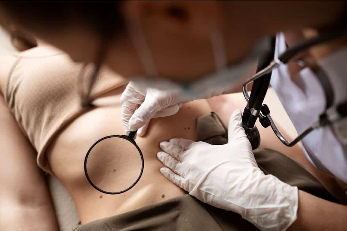
When Professional Mole Assessment Is Necessary
Seek immediate professional assessment if you notice:
- Any new mole appearing after age 30
- A mole that follows the ABCDE criteria
- A mole that bleeds, itches, or causes pain
- A rapidly growing mole
- A mole that looks different from others (“ugly duckling” sign)
Family history matters too. The NHS Genomics Education programme emphasises that those with multiple melanomas or atypical moles combined with family history face higher risks and need more frequent monitoring.
High UV exposure can cause temporary changes in moles that mimic melanoma, making professional assessment crucial rather than self-diagnosis.
Safe Mole Removal Procedures
When professional removal is recommended, several methods might be used:
Surgical excision – The entire mole is cut out along with a small margin of surrounding skin. This allows for complete histological analysis.
Shave excision – The mole is shaved off level with the skin. This works well for raised moles but doesn’t allow examination of deeper tissues.
Punch biopsy – A small circular blade removes a deeper core of skin. This provides depth without removing as much surface area.
Each method has specific applications based on the mole’s characteristics and location. The gold standard remains surgical excision with histological analysis.
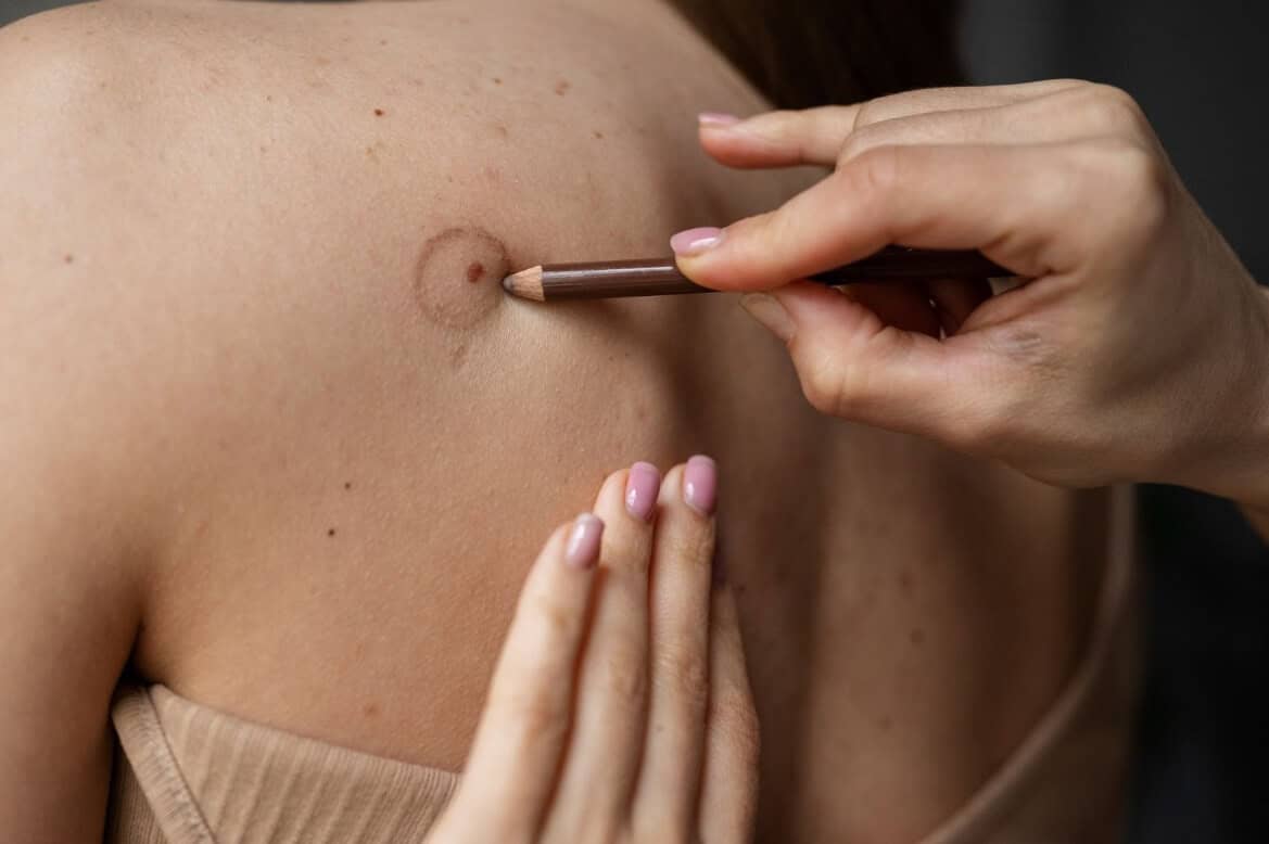
The Dangers of DIY Mole Removal
At-home mole removal carries serious risks that far outweigh any convenience:
- Without biopsy, you might miss melanoma diagnosis, allowing cancer to spread undetected
- Improper technique can cause infections, excessive scarring, or keloids
- DIY creams and lotions often cause skin damage and can increase scarring
- Attempting to cut out moles can spread cancerous cells if the mole is malignant
The Skin Cancer Foundation strongly warns against DIY removal, noting cases where delayed diagnosis from improper home treatment led to preventable metastasis.
How Aventus Clinic Approaches Mole Assessment
At Aventus Clinic, we take a comprehensive approach to mole checking service. Our process includes:

For suspicious moles, we offer safe removal procedures performed by experienced professionals, ensuring proper handling and testing of all tissue samples.
Long-term Skin Health After Mole Removal
After professional mole removal, long-term skin health involves:
Monitoring the healing site: Some redness and slight scarring are normal but should improve over time.
Regular skin checks: Having one suspicious mole increases the likelihood of developing others.
Sun protection: UV exposure remains the biggest risk factor for developing melanoma.
Follow-up appointments: These ensure proper healing and catch any new concerns early.
Conclusion
The truth about mole removal and cancer is clear: professional assessment and removal do not cause cancer. In fact, they often prevent it. The real danger lies in ignoring concerning changes or attempting DIY removal.
If you have a mole that worries you, don’t wait. Book a free online assessment with our specialists today. Early intervention could save your life.
Remember, most moles are harmless. But knowing which ones aren’t makes all the difference.
- Sources
- https://www.ncbi.nlm.nih.gov/books/NBK470409/
- https://www.skincancer.org/risk-factors/atypical-moles/
- https://pmc.ncbi.nlm.nih.gov/articles/PMC5930388/
- https://www.genomicseducation.hee.nhs.uk/genotes/in-the-clinic/presentation-patient-with-cutaneous-melanoma/
- https://www.skincancer.org/blog/diy-donts-why-at-home-mole-removal-is-a-bad-idea/


 CALL US
 WHATSAPP