Overview
What is cherry angioma?
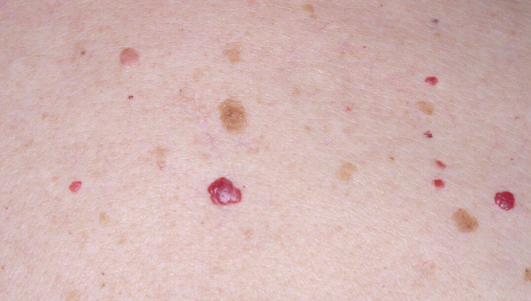
Cherry angiomas are typically small, usually measuring between about 1 mm and 5 mm in diameter, although they can occasionally be slightly larger. They often develop on the trunk (chest and abdomen), arms, legs or shoulders, and may increase in number with age.
Cherry angiomas are benign vascular proliferations, meaning they result from clusters of capillaries (tiny blood vessels) just beneath the surface of the skin. They are not contagious, not harmful to general health and do not have malignant potential under normal circumstances.
Symptoms and Causes
Symptoms
Cherry angiomas are usually:
- Bright red, purple or maroon spots on the skin
- Smooth and slightly raised above the skin surface
- Painless and asymptomatic in most cases
They are typically discovered during routine skin checks and rarely cause discomfort. However, they may bleed if scratched, rubbed or injured due to their blood-filled nature.
Causes
The exact cause of cherry angiomas is unknown, but several factors have been linked with their development:
- Age: They become more common after age 30 and increase in prevalence with advancing age.
- Genetic factors: There may be a hereditary component in some families.
- Hormonal influences: They may be seen more frequently during pregnancy.
- Chemical exposures: Certain chemicals can be associated with their appearance.
Although they are often referred to as “senile angiomas,” they can also occur in younger adults, and small numbers may be seen even in adolescence.
Sudden appearance of multiple lesions
In rare cases, a sudden appearance of multiple lesions can occur. This is known as eruptive cherry angiomas. If many new angiomas develop over a short period, it is sensible to seek medical advice to rule out uncommon underlying causes.
Diagnosis and Tests
Diagnosis
Cherry angiomas are usually diagnosed through clinical examination by a healthcare professional based on their characteristic appearance. No routine tests are needed for diagnosis.
In uncertain cases, such as when a lesion has unusual features, changes in appearance or causes concern, further assessment may include:
- Dermoscopy, which typically shows well-defined red or purple lacunae separated by thin septa
- Biopsy, if there is uncertainty or concern about melanoma or another condition
Differential Diagnosis
Although cherry angiomas are benign, they can sometimes resemble other red-coloured lesions, including:
- Spider angiomas
- Pyogenic granulomas
- Angiokeratomas
- Amelanotic melanoma
If a lesion changes rapidly, bleeds often without injury, or has irregular features, further assessment is advisable to rule out other conditions.
Management and Treatment
When treatment is needed?
Most cherry angiomas do not require medical treatment, as they are asymptomatic and pose no risk to health. People may choose treatment for cosmetic reasons or if a lesion:
- Bleeds frequently due to irritation or friction
- Causes discomfort
- Appears in a highly visible area
Treatment options
Removal should always be performed by a trained clinician. Common methods include:
- Electrodesiccation: An electric needle is used to destroy the lesion.
- Cryotherapy: Liquid nitrogen is applied to freeze the lesion.
- Laser therapy: Laser light targets the blood vessels to remove the angioma.
These procedures are normally quick and can often be done in a dermatology clinic with minimal downtime. Slight scarring is a possible side effect.
It is important not to attempt removal at home, as doing so can cause bleeding, infection and scarring.
Outlook/Prognosis
Cherry angiomas have an excellent prognosis. They are benign and not associated with skin cancer. They often remain stable for many years once they form and may increase in number with age.
Removing a cherry angioma does not guarantee that new lesions will not appear; recurrence or development of new angiomas is common, especially as a person ages.
Prevention
There is no known way to prevent cherry angiomas entirely because their direct cause is not understood. Avoiding exposure to certain chemicals known to be associated with angioma formation may reduce risk, but this is not guaranteed.
Protecting skin from injury may help prevent bleeding in existing lesions, and regular skin checks help ensure any changes are evaluated promptly.