

Cherry angiomas are one of the most common benign skin growths seen in adults. They often appear as small red, purple or burgundy spots on the trunk, arms or shoulders.
- Dr Sharon Crichlow
- Reading Time: 10 Mins
Many people notice them increasing with age and naturally wonder whether they will disappear without treatment. In most cases, cherry angiomas do not go away on their own. Once they form, they usually remain unless removed by a healthcare professional.
This article explains what cherry angiomas are, why they persist, when to seek medical advice, and addresses common concerns.
Key Takeaways
- Cherry angiomas are common, benign vascular skin growths that usually persist once they develop. They do not typically go away on their own and may slowly increase in number with age.
- A sudden appearance of multiple lesions is often harmless, but rapid changes, unusual features or associated symptoms should be assessed by a healthcare professional to confirm the diagnosis.
- Cherry angiomas are not contagious and cannot be prevented through diet or lifestyle changes. Removal is optional and should only be carried out by a qualified clinician if needed for cosmetic reasons or repeated bleeding.
Table of Contents
What Are Cherry Angiomas?
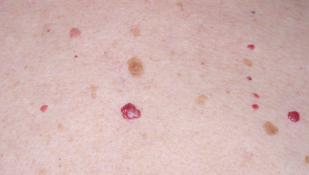
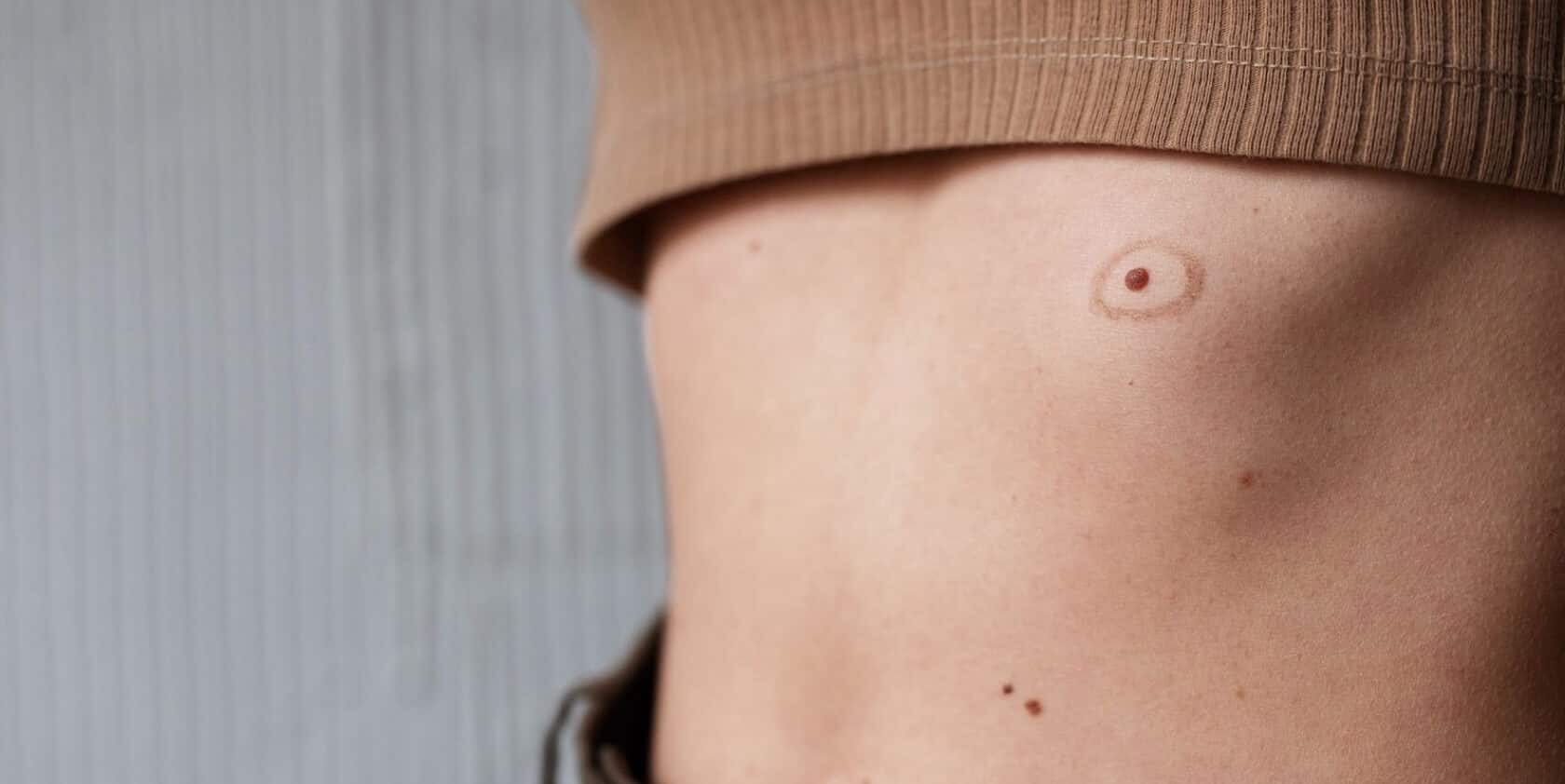
Cherry angiomas are harmless growths made up of clusters of tiny dilated blood vessels known as capillaries. They are sometimes referred to as Campbell de Morgan spots or red moles.
They typically appear bright red, dark red or purple in colour. Most are small, measuring between 1 and 5 millimetres, although some may enlarge over time. They are often smooth, round or dome shaped and may be flat or slightly raised.
These lesions most commonly develop on the chest, abdomen, back, shoulders and upper arms. They are extremely common, particularly in adults over the age of 30, and the number of lesions often increases gradually with ageing. Cherry angiomas are not infectious and are not caused by poor hygiene.

Do Cherry Angiomas Go Away on Their Own?
Cherry angiomas that develop in adulthood do not usually disappear without treatment.
Unlike infantile haemangiomas, which are vascular birthmarks seen in babies that often shrink during childhood, cherry angiomas are stable clusters of blood vessels. Once they have formed, there is no recognised natural biological process that causes them to regress.
Some lesions may darken slightly or flatten over many years, but true spontaneous disappearance is uncommon. For most individuals, cherry angiomas remain stable or gradually increase in number over time.
Why Do Cherry Angiomas Persist?
Cherry angiomas form due to a benign increase in small blood vessels within the superficial layer of the skin. Although the precise cause is not fully understood, ageing is considered a major contributing factor. Genetic predisposition may also play a role.
Because these lesions are structural collections of capillaries rather than temporary inflammatory changes, they tend to persist once formed. Over time they may remain unchanged, enlarge slowly, or increase in number. Their presence does not indicate infection or malignancy.
Can Cherry Angiomas Appear Suddenly in Large Numbers?
Some people notice multiple cherry angiomas appearing over what seems to be a short period. In many cases, this reflects gradual development that becomes more noticeable over time.
For most individuals, the appearance of several lesions is still consistent with benign, age related skin change.
However, a sudden eruption of numerous angiomas, particularly if accompanied by other unexplained symptoms, should be assessed by a healthcare professional. Although uncommon, widespread eruptive lesions may occasionally be associated with medication effects or underlying medical conditions. Clinical evaluation helps confirm that the lesions are typical cherry angiomas.
Is a Sudden Eruption Something to Worry About?
Most cherry angiomas are harmless and require no treatment.
Medical advice should be sought if many lesions appear rapidly, if there are additional unexplained symptoms, or if the lesions look atypical. A clinician can usually confirm the diagnosis through visual examination and, if needed, dermoscopy.
Early assessment is advisable whenever there is uncertainty.
How Do Cherry Angiomas Differ From Other Red Skin Lesions?
Several skin conditions can resemble cherry angiomas, and distinguishing between them is important.
- Cherry angiomas are typically well defined, bright red to purple in colour, and round or dome shaped. They are often slightly raised and may bleed if scratched because they consist of small blood vessels.
- Petechiae appear as very small, flat red or purple dots that do not blanch when pressed. They are caused by bleeding under the skin rather than dilated surface blood vessels and may be associated with medical conditions affecting clotting or platelet function.
- Spider angiomas consist of a central red spot with fine blood vessels radiating outward. They often blanch with pressure and are commonly seen during pregnancy or in association with certain liver conditions.
- Pyogenic granulomas present as rapidly growing red lumps that are soft and prone to bleeding. They often develop after minor injury and tend to enlarge more quickly than cherry angiomas.
- Any red lesion that does not clearly match the appearance of a typical cherry angioma should be examined by a healthcare professional.
Comparison of Cherry Angiomas and Other Red Skin Lesions
| Feature | Cherry Angioma | Petechiae | Spider Angioma | Pyogenic Granuloma |
|---|---|---|---|---|
| Typical appearance | Bright red to purple, round or dome shaped | Very small red or purple dots | Central red spot with fine radiating vessels | Red lump, often smooth and shiny |
| Surface level | Slightly raised or dome shaped | Flat | Usually flat central spot with visible vessel pattern | Raised and often protruding |
| Size | Usually 1 to 5 mm, may enlarge slowly | Pinpoint sized | Small central lesion with surrounding fine vessels | Can grow rapidly and become larger than 5 mm |
| Blanching with pressure | May partially blanch | Does not blanch | Usually blanches when pressed | May bleed rather than blanch |
| Growth pattern | Slow growth or stable over time | Appear suddenly, often multiple | May appear gradually | Rapid growth over weeks |
| Cause | Cluster of dilated capillaries | Bleeding under the skin | Dilated superficial blood vessels | Reactive vascular overgrowth, often after injury |
| Bleeding tendency | May bleed if scratched | Do not bleed externally | Rarely bleed | Frequently bleed easily |
| Associated factors | Common with ageing | May relate to clotting disorders or infection | Pregnancy, liver conditions | Minor trauma or skin irritation |
| Serious concern level | Benign | May require medical assessment | Usually benign | Benign but often treated due to bleeding |
Important: If a red skin lesion changes rapidly, grows quickly, becomes irregular, ulcerates, or does not clearly match the typical features of a cherry angioma, medical assessment is recommended to confirm the diagnosis.
Can Cherry Angiomas Be Mistaken for Skin Cancer?
Cherry angiomas are benign and do not develop into melanoma. However, certain skin cancers, including amelanotic melanoma, can appear pink or red and may resemble vascular lesions. A lesion should be assessed if it changes rapidly, develops irregular borders, ulcerates, grows steadily, or appears asymmetrical. Prompt medical evaluation ensures accurate diagnosis and appropriate management.
Why Do Some People Develop Cherry Angiomas at a Younger Age?
Although more common after the age of 30, cherry angiomas can develop in younger adults. Genetic predisposition, natural variation in vascular development and hormonal influences may contribute. In most cases, there is no identifiable cause and the lesions remain harmless. The presence of cherry angiomas at a younger age does not usually indicate underlying disease.
Can Cherry Angiomas Be Prevented?
There is no proven way to prevent cherry angiomas. They are not reliably linked to diet, skincare products, hygiene or general lifestyle habits. Rare associations have been described with certain chemical exposures or medical conditions, but for the vast majority of people they represent a normal age related change in the skin.
General skin care measures, including appropriate sun protection, are important for overall skin health but have not been shown to prevent cherry angiomas specifically.
Is It Safe to Remove Cherry Angiomas at Home?
Home removal is not recommended. Because cherry angiomas are composed of blood vessels, attempts to cut, burn or puncture them can lead to significant bleeding, infection, scarring and delayed diagnosis of a more serious condition.
Over the counter devices and internet methods are not substitutes for medical assessment. Removal should only be performed by a qualified healthcare professional.
When Is Treatment Appropriate?
Treatment is usually optional and considered for cosmetic reasons or if the lesion bleeds frequently or becomes irritated by clothing or shaving. Medical removal methods include laser therapy, electrocautery, cryotherapy and minor surgical excision. These procedures are typically quick and carried out in an outpatient setting.
Conclusion
Cherry angiomas are common benign vascular growths that increase with age. They do not usually go away on their own and tend to persist once formed. Although generally harmless, any lesion that changes in appearance, grows rapidly or appears suddenly in large numbers should be assessed by a healthcare professional. Removal is optional and should only be carried out by trained clinicians.
FAQs
Do cherry angiomas grow over time?
Cherry angiomas can increase slightly in size over time, although many remain stable for years. They may also become darker in colour. Growth is usually slow. Rapid enlargement should be assessed to confirm the diagnosis.
Can cherry angiomas bleed heavily?
Because cherry angiomas are made up of small blood vessels, they can bleed if scratched, cut or traumatised. The bleeding may appear significant due to the vascular nature of the lesion, but it is usually manageable with firm pressure. Persistent or recurrent bleeding should be reviewed by a healthcare professional.
Are cherry angiomas painful?
Cherry angiomas are typically painless. They may become tender only if irritated by friction, shaving or injury. Pain without obvious trauma is unusual and should be assessed.
Can cherry angiomas appear on the face?
Yes, although they are most common on the trunk, cherry angiomas can appear on the face, scalp and other areas of the body. Facial lesions may cause cosmetic concern, but they are usually harmless.
Are cherry angiomas linked to high blood pressure?
There is no strong evidence to suggest that cherry angiomas are caused by or directly linked to high blood pressure. They are primarily associated with ageing and genetic factors.
Can medications cause cherry angiomas?
In most cases, cherry angiomas occur independently of medication use. Rarely, certain drugs have been reported in association with eruptive angiomas. If multiple lesions appear shortly after starting a new medication, medical advice should be sought.
Are cherry angiomas contagious?
No. Cherry angiomas are not contagious. They cannot be spread through skin contact and are not caused by infection.
Do cherry angiomas indicate an underlying internal disease?
For the vast majority of people, cherry angiomas are a benign age related skin change and do not indicate internal disease. If lesions appear suddenly in large numbers or are accompanied by other symptoms, medical assessment is appropriate to exclude rare underlying causes.
Can cherry angiomas turn black?
Cherry angiomas may darken over time and can appear deep red, purple or almost black, particularly if thrombosed, which means a small clot has formed within the lesion. A sudden colour change should be reviewed to confirm it remains a benign angioma.
How are cherry angiomas confirmed by a doctor?
Cherry angiomas are usually diagnosed through clinical examination. A doctor may use dermoscopy, a handheld magnifying device with light, to examine the vascular pattern. Biopsy is rarely required unless the diagnosis is uncertain.
Not sure if your red skin lesion is a cherry angioma? Get expert advice from our dermatology team with a free online skin assessment today.
- Medical Disclaimer
This information is for general guidance only and does not replace medical advice. If you are concerned about a skin lesion, request a personalised assessment from our qualified dermatology team through our free online service.
 CALL US
 WHATSAPP